1932

Abstract

Cerebrospinal fluid (CSF) is not stagnant but displays fascinating oscillatory flow patterns inside the ventricular system and reversing fluid exchange between the cranial vault and spinal compartment. This review provides an overview of the current knowledge of pulsatile CSF motion. Observations contradicting classical views about its bulk production and clearance are highlighted. A clinical account of diseases of abnormal CSF flow dynamics, including hydrocephalus, syringomyelia, Chiari malformation type 1, and pseudotumor cerebri, is also given. We survey medical imaging modalities used to observe intracranial dynamics in vivo. Additionally, we assess the state of the art in predictive models of CSF dynamics. The discussion addresses open questions regarding CSF dynamics as they relate to the understanding and management of diseases.

Loading

Article metrics loading...

/content/journals/10.1146/annurev-fluid-122414-034321
2016-01-03
2024-04-18
Loading full text...

Full text loading...

/deliver/fulltext/fluid/48/1/annurev-fluid-122414-034321.html?itemId=/content/journals/10.1146/annurev-fluid-122414-034321&mimeType=html&fmt=ahah

Literature Cited

  1. Algin O, Turkbey B. 2012. Evaluation of aqueductal stenosis by 3D sampling perfection with application-optimized contrasts using different flip angle evolutions sequence: preliminary results with 3T MR imaging. Am. J. Neuroradiol. 33:740–46 [Google Scholar]
  2. Alperin N, Hushek SG, Lee SH, Sivaramakrishnan A, Lichtor T. 2005. MRI study of cerebral blood flow and CSF flow dynamics in an upright posture: the effect of posture on the intracranial compliance and pressure. Intracranial Pressure and Brain Monitoring XII WS Poon, MTV Chan, KYC Goh, JMK Lam, SCP Ng, et al. 177–81 New York: Springer [Google Scholar]
  3. Ambarki K, Lindqvist T, Wåhlin A, Petterson E, Warntjes MJB. et al. 2012. Evaluation of automatic measurement of the intracranial volume based on quantitative MR imaging. Am. J. Neuroradiol. 33:1951–56 [Google Scholar]
  4. Amin-Hanjani S, Shin JH, Zhao M, Du X, Charbel FT. 2007. Evaluation of extracranial–intracranial bypass using quantitative magnetic resonance angiography. J. Neurosurg. 106:291–98 [Google Scholar]
  5. Armonda RA, Citrin CM, Foley KT, Ellenbogen RG. 1994. Quantitative cine-mode magnetic resonance imaging of Chiari I malformations: an analysis of cerebrospinal fluid dynamics. Neurosurgery 35:214–23 discussion 223–24 [Google Scholar]
  6. Attique M, Gilanie G, Hafeez-Ullah, Mehmood MS, Naweed MS. et al. 2012. Colorization and automated segmentation of human T2 MR brain images for characterization of soft tissues. PLOS ONE 7:e33616 [Google Scholar]
  7. Aubin ML, Vignaud J, Jardin C, Bar D. 1981. Computed tomography in 75 clinical cases of syringomyelia. Am. J. Neuroradiol. 2:199–204 [Google Scholar]
  8. Auer LM, Ishiyama N. 1986. Cerebrovascular response to elevated intracranial pressure. Intracranial Pressure VI PJD Miller, PGM Teasdale, PJO Rowan, PSL Galbraith, PAD Mendelow 399–403 New York: Springer [Google Scholar]
  9. Balédent O, Gondry-Jouet C, Meyer M-E, De Marco G, Le Gars D. et al. 2004. Relationship between cerebrospinal fluid and blood dynamics in healthy volunteers and patients with communicating hydrocephalus. Invest. Radiol. 39:45–55 [Google Scholar]
  10. Battal B, Kocaoglu M, Bulakbasi N, Husmen G, Tuba Sanal H, Tayfun C. 2011. Cerebrospinal fluid flow imaging by using phase-contrast MR technique. Br. J. Radiol. 84:758–65 [Google Scholar]
  11. Bauer AM, Amin-Hanjani S, Alaraj A, Charbel FT. 2009. Quantitative magnetic resonance angiography in the evaluation of the subclavian steal syndrome: report of 5 patients. J. Neuroimaging 19:250–52 [Google Scholar]
  12. Bergstrand G, Bergström M, Nordell B, Ståhlberg F, Ericsson A. et al. 1985. Cardiac gated MR imaging of cerebrospinal fluid flow. J. Comput. Assist. Tomogr. 9:1003–6 [Google Scholar]
  13. Bering EA. 1952. Water exchange of central nervous system and cerebrospinal fluid. J. Neurosurg. 9:275–87 [Google Scholar]
  14. Bernards CM. 2000. Rostral spread of epidural morphine: the expected and the unexpected. Anesthesiology 92:299–301 [Google Scholar]
  15. Bernards CM, Shen DD, Sterling ES, Adkins JE, Risler L. et al. 2003. Epidural, cerebrospinal fluid, and plasma pharmacokinetics of epidural opioids (part 1): differences among opioids. Anesthesiology 99:455–65 [Google Scholar]
  16. Bhadelia RA, Bogdan AR, Wolpert SM. 1995. Analysis of cerebrospinal fluid flow waveforms with gated phase-contrast MR velocity measurements. Am. J. Neuroradiol. 16:389–400 [Google Scholar]
  17. Bhadelia RA, Madan N, Zhao Y, Wagshul ME, Heilman C. et al. 2013. Physiology-based MR imaging assessment of CSF flow at the foramen magnum with a valsalva maneuver. Am. J. Neuroradiol. 34:1857–62 [Google Scholar]
  18. Bilston LE, Fletcher DF, Brodbelt AR, Stoodley MA. 2003. Arterial pulsation-driven cerebrospinal fluid flow in the perivascular space: a computational model. Comput. Methods Biomech. Biomed. Eng. 6:235–41 [Google Scholar]
  19. Bilston LE, Stoodley MA, Fletcher DF. 2009. The influence of the relative timing of arterial and subarachnoid space pulse waves on spinal perivascular cerebrospinal fluid flow as a possible factor in syrinx development. J. Neurosurg. 112:808–13 [Google Scholar]
  20. Bolash R, Udeh B, Saweris Y, Guirguis M, Dalton JE. et al. 2015. Longevity and cost of implantable intrathecal drug delivery systems for chronic pain management: a retrospective analysis of 365 patients. Neuromodul. Technol. Neural Interface 18:150–56 [Google Scholar]
  21. Bottan S, Poulikakos D, Kurtcuoglu V. 2012. Phantom model of physiologic intracranial pressure and cerebrospinal fluid dynamics. IEEE Trans. Biomed. Eng. 59:1532–38 [Google Scholar]
  22. Bottan S, Schmid Daners M, de Zelicourt D, Fellner N, Poulikakos D, Kurtcuoglu V. 2013. Assessment of intracranial dynamics in hydrocephalus: effects of viscoelasticity on the outcome of infusion tests. J. Neurosurg. 119:1511–19 [Google Scholar]
  23. Boulton M, Flessner M, Armstrong D, Mohamed R, Hay J, Johnston M. 1999. Contribution of extracranial lymphatics and arachnoid villi to the clearance of a CSF tracer in the rat. Am. J. Physiol. 276:R818–23 [Google Scholar]
  24. Bouma GJ, Muizelaar JP, Bandoh K, Marmarou A. 1992. Blood pressure and intracranial pressure-volume dynamics in severe head injury: relationship with cerebral blood flow. J. Neurosurg. 77:15–19 [Google Scholar]
  25. Brisman JL, Pile-Spellman J, Konstas AA. 2012. Clinical utility of quantitative magnetic resonance angiography in the assessment of the underlying pathophysiology in a variety of cerebrovascular disorders. Eur. J. Radiol. 81:298–302 [Google Scholar]
  26. Brodbelt A, Stoodley M. 2007. CSF pathways: a review. Br. J. Neurosurg. 21:510–20 [Google Scholar]
  27. Bruel BM, Engle MP, Rauck RL, Weber TJ, Kapural L. 2013. Intrathecal drug delivery for control of pain. Comprehensive Treatment of Chronic Pain by Medical, Interventional, and Integrative Approaches TR Deer, MS Leong, A Buvanendran, V Gordin, PS Kim, et al. 637–48 New York: Springer [Google Scholar]
  28. Buishas J, Gould IG, Linninger AA. 2014. A computational model of cerebrospinal fluid production and reabsorption driven by Starling forces. Croat. Med. J. 55:481–97 [Google Scholar]
  29. Bulat M, Klarica M. 2011. Recent insights into a new hydrodynamics of the cerebrospinal fluid. Brain Res. Rev. 65:99–112 [Google Scholar]
  30. Bulat M, Lupret V, Orehković D, Klarica M. 2008. Transventricular and transpial absorption of cerebrospinal fluid into cerebral microvessels. Coll. Antropol. 32:Suppl. 143–50 [Google Scholar]
  31. Bunck AC, Kröger JR, Jüttner A, Brentrup A, Fiedler B. et al. 2011. Magnetic resonance 4D flow characteristics of cerebrospinal fluid at the craniocervical junction and the cervical spinal canal. Eur. Radiol. 21:1788–96 [Google Scholar]
  32. Bunck AC, Kröger JR, Jüttner A, Brentrup A, Fiedler B. et al. 2012. Magnetic resonance 4D flow analysis of cerebrospinal fluid dynamics in Chiari I malformation with and without syringomyelia. Eur. Radiol. 22:1860–70 [Google Scholar]
  33. Cahan LD, Bentson JR. 1982. Considerations in the diagnosis and treatment of syringomyelia and the Chiari malformation. J. Neurosurg. 57:24–31 [Google Scholar]
  34. Cai J, Chang Z, Wang Z, Segars WP, Yin F-F. 2011. Four-dimensional magnetic resonance imaging (4D-MRI) using image-based respiratory surrogate: a feasibility study. Med. Phys. 38:6384–94 [Google Scholar]
  35. Calias P, Banks WA, Begley D, Scarpa M, Dickson P. 2014. Intrathecal delivery of protein therapeutics to the brain: a critical reassessment. Pharmacol. Ther. 144:114–22 [Google Scholar]
  36. Calias P, Papisov M, Pan J, Savioli N, Belov VV. et al. 2012. CNS penetration of intrathecal-lumbar idursulfase in the monkey, dog and mouse: implications for neurological outcomes of lysosomal storage disorder. PLOS ONE 7:e30341 [Google Scholar]
  37. Carare RO, Bernardes-Silva M, Newman TA, Page AM, Nicoll JAR. et al. 2008. Solutes, but not cells, drain from the brain parenchyma along basement membranes of capillaries and arteries: significance for cerebral amyloid angiopathy and neuroimmunology. Neuropathol. Appl. Neurobiol. 34:131–44 [Google Scholar]
  38. Chang HS, Nakagawa H. 2003. Hypothesis on the pathophysiology of syringomyelia based on simulation of cerebrospinal fluid dynamics. J. Neurol. Neurosurg. Psychiatry 74:344–47 [Google Scholar]
  39. Charbel FT, Zhao M, Amin-Hanjani S, Hoffman W, Du X, Clark ME. 2004. A patient-specific computer model to predict outcomes of the balloon occlusion test. J. Neurosurg. 101:977–88 [Google Scholar]
  40. Cheng S, Fletcher D, Hemley S, Stoodley M, Bilston L. 2014. Effects of fluid structure interaction in a three dimensional model of the spinal subarachnoid space. J. Biomech. 47:2826–30 [Google Scholar]
  41. Cheng S, Stoodley MA, Wong J, Hemley S, Fletcher DF, Bilston LE. 2012. The presence of arachnoiditis affects the characteristics of CSF flow in the spinal subarachnoid space: a modelling study. J. Biomech. 45:1186–91 [Google Scholar]
  42. Clarke EC, Stoodley MA, Bilston LE. 2013. Changes in temporal flow characteristics of CSF in Chiari malformation Type I with and without syringomyelia: implications for theory of syrinx development. J. Neurosurg. 118:1135–40 [Google Scholar]
  43. Cohen B, Voorhees A, Vedel S, Wei T. 2009. Development of a theoretical framework for analyzing cerebrospinal fluid dynamics. Cerebrospinal Fluid Res. 6:12 [Google Scholar]
  44. Cousins J, Haughton V. 2009. Motion of the cerebellar tonsils in the foramen magnum during the cardiac cycle. Am. J. Neuroradiol. 30:1587–88 [Google Scholar]
  45. Cserr HF. 1971. Physiology of the choroid plexus. Physiol. Rev. 51:273–311 [Google Scholar]
  46. Detrembleur C, Plaghki L. 2000. Quantitative assessment of intrathecally administered baclofen in spasticity. Arch. Phys. Med. Rehabil. 81:279–84 [Google Scholar]
  47. Dreha-Kulaczewski S, Joseph AA, Merboldt K-D, Ludwig H-C, Gärtner J, Frahm J. 2015. Inspiration is the major regulator of human CSF flow. J. Neurosci. 35:2485–91 [Google Scholar]
  48. Ducoffe AR, Konstas AA, Pile-Spellman J, Brisman JL. 2012. Clinical applications of quantitative MRA in neurovascular practice. Magnetic Resonance Angiography: Basics to Future W Shabana. Rijeka, Croat.: InTech. doi: 10.5772/29874
  49. Dykstra D, Stuckey M, DesLauriers L, Chappuis D, Krach L. 2007. Intrathecal baclofen in the treatment of spasticity. Operative Neuromodulation DE Sakas, BA Simpson, ES Krames 163–71 New York: Springer [Google Scholar]
  50. Edsbagge M, Tisell M, Jacobsson L, Wikkelso C. 2004. Spinal CSF absorption in healthy individuals. Am. J. Physiol. 287:R1450–55 [Google Scholar]
  51. Egnor M, Rosiello A, Zheng L. 2001. A model of intracranial pulsations. Pediatr. Neurosurg. 35:284–98 [Google Scholar]
  52. Ehlers W, Wagner A. 2013. Constitutive and computational aspects in tumor therapies of multiphasic brain tissue. Computer Models in Biomechanics GA Holzapfel, E Kuhl 263–76 New York: Springer [Google Scholar]
  53. Ehlers W, Wagner A. 2015. Multi-component modelling of human brain tissue: a contribution to the constitutive and computational description of deformation, flow and diffusion processes with application to the invasive drug-delivery problem. Comput. Methods Biomech. Biomed. Eng. 18:861–79 [Google Scholar]
  54. Enzmann DR, Pelc NJ. 1992. Brain motion: measurement with phase-contrast MR imaging. Radiology 185:653–60 [Google Scholar]
  55. Enzmann DR, Pelc NJ. 1993. Cerebrospinal fluid flow measured by phase-contrast cine MR. AJNR Am. J. Neuroradiol. 14:1301–7 discussion 1309–10 [Google Scholar]
  56. Esfahani D, Stevenson M, Moss H, Amin-Hanjani S, Aletich V. et al. 2014. O-011 quantitative MRA is correlated with intravascular pressures before and after venous sinus stenting: implications for treatment and monitoring. J. Neurointerv. Surg. 6:Suppl. 1A6–7 [Google Scholar]
  57. Fernández E, Graña M, Villanúa J. 2011. High resolution segmentation of CSF on phase contrast MRI. New Challenges on Bioinspired Applications JM Ferrández, JR Álvarez, F de la Paz, FJ Toledo 96–103 New York: Springer [Google Scholar]
  58. Freund M, Adwan M, Kooijman H, Heiland S, Thomsen M. et al. 2001. Measurement of CSF flow in the spinal canal using MRI with an optimized MRI protocol: experimental and clinical studies. RöFo 173:306–14 [Google Scholar]
  59. Friedman DI, Jacobson DM. 2002. Diagnostic criteria for idiopathic intracranial hypertension. Neurology 59:1492–95 [Google Scholar]
  60. Galarza M, Giménez Á, Pellicer O, Valero J, Amigó JM. 2014. New designs of ventricular catheters for hydrocephalus by 3-D computational fluid dynamics. Childs Nerv. Syst. 31:37–48 [Google Scholar]
  61. Galarza M, Giménez Á, Valero J, Pellicer OP, Amigó JM. 2013. Computational fluid dynamics of ventricular catheters used for the treatment of hydrocephalus: a 3D analysis. Childs Nerv. Syst. 30:105–16 [Google Scholar]
  62. Galarza M, Giménez Á, Valero J, Pellicer O, Martínez-Lage JF, Amigó JM. 2015. Basic cerebrospinal fluid flow patterns in ventricular catheters prototypes. Childs Nerv. Syst. 31:873–84 [Google Scholar]
  63. George TM, Higginbotham NH. 2011. Defining the signs and symptoms of Chiari malformation type I with and without syringomyelia. Neurol. Res. 33:240–46 [Google Scholar]
  64. Ghaffari M, Hsu C, Linninger A. 2015. Automatic reconstruction and generation of structured hexahedral mesh for non-planar bifurcations in vascular networks Presented at 12th Int. Symp. Process Syst. Eng., 25th Eur. Symp. Comput. Aided Process Eng, Copenhagen
  65. Gould IG, Linninger AA. 2015. Hematocrit distribution and tissue oxygenation in large microcirculatory networks. Microcirculation 22:1–18 [Google Scholar]
  66. Gracies J-M, Nance P, Elovic E, McGuire J, Simpson DM. 1997. Traditional pharmacological treatments for spasticity. Part II: general and regional treatments. Muscle Nerve 20:Suppl. 6S92–120 [Google Scholar]
  67. Greitz D. 2004. Radiological assessment of hydrocephalus: new theories and implications for therapy. Neurosurg. Rev. 27:145–65 [Google Scholar]
  68. Greitz D, Hannerz J, Rähn T, Bolander H, Ericsson A. 1994. MR imaging of cerebrospinal fluid dynamics in health and disease on the vascular pathogenesis of communicating hydrocephalus and benign intracranial hypertension. Acta Radiol. 35:204–11 [Google Scholar]
  69. Gupta S, Poulikakos D, Kurtcuoglu V. 2008. Analytical solution for pulsatile viscous flow in a straight elliptic annulus and application to the motion of the cerebrospinal fluid. Phys. Fluids 20:093607 [Google Scholar]
  70. Gupta S, Soellinger M, Boesiger P, Poulikakos D, Kurtcuoglu V. 2009. Three-dimensional computational modeling of subject-specific cerebrospinal fluid flow in the subarachnoid space. J. Biomech. Eng. 131:021010 [Google Scholar]
  71. Gupta S, Soellinger M, Grzybowski DM, Boesiger P, Biddiscombe J. et al. 2010. Cerebrospinal fluid dynamics in the human cranial subarachnoid space: an overlooked mediator of cerebral disease. I. Computational model. J. R. Soc. Interface 7:1195–204 [Google Scholar]
  72. Hadaczek P, Yamashita Y, Mirek H, Tamas L, Bohn MC. et al. 2006. The “perivascular pump” driven by arterial pulsation is a powerful mechanism for the distribution of therapeutic molecules within the brain. Mol. Ther. 14:69–78 [Google Scholar]
  73. Hadzri EA, Osman K, Kadir MRA, Aziz AA. 2011. Computational investigation on CSF flow analysis in the third ventricle and aqueduct of Sylvius. IIUM Eng. J. 12:132–41 [Google Scholar]
  74. Haj-Yasein NN, Vindedal GF, Eilert-Olsen M, Gundersen GA, Skare Ø. et al. 2011. Glial-conditional deletion of aquaporin-4 (Aqp4) reduces blood-brain water uptake and confers barrier function on perivascular astrocyte endfeet. PNAS 108:17815–20 [Google Scholar]
  75. Hakim S. 1970. Biomechanics of hydrocephalus. Acta Neurol. Latinoam. 1:Suppl. 1169–94 [Google Scholar]
  76. Hakim S, Venegas JG, Burton JD. 1976. The physics of the cranial cavity, hydrocephalus and normal pressure hydrocephalus: mechanical interpretation and mathematical model. Surg. Neurol. 5:187–210 [Google Scholar]
  77. Haller FR, Low FN. 1971. The fine structure of the peripheral nerve root sheath in the subarachnoid space in the rat and other laboratory animals. Am. J. Anat. 131:1–19 [Google Scholar]
  78. Haslam M, Zamir M. 1998. Pulsatile flow in tubes of elliptic cross sections. Ann. Biomed. Eng. 26:780–87 [Google Scholar]
  79. Hebb AO, Cusimano MD. 2001. Idiopathic normal pressure hydrocephalus: a systematic review of diagnosis and outcome. Neurosurgery 49:1166–84 discussion 1184–86 [Google Scholar]
  80. Helgeland A, Mardal K-A, Haughton V, Reif BAP. 2014. Numerical simulations of the pulsating flow of cerebrospinal fluid flow in the cervical spinal canal of a Chiari patient. J. Biomech. 47:1082–90 [Google Scholar]
  81. Hernández-Mier Y, Blondel WCPM, Daul C, Wolf D, Guillemin F. 2010. Fast construction of panoramic images for cystoscopic exploration. Comput. Med. Imaging Graph. 34:579–92 [Google Scholar]
  82. Hettiarachchi HDM, Hsu Y, Harris TJ, Penn R, Linninger AA. 2011. The effect of pulsatile flow on intrathecal drug delivery in the spinal canal. Ann. Biomed. Eng. 39:2592–602 [Google Scholar]
  83. Hladky SB, Barrand MA. 2014. Mechanisms of fluid movement into, through and out of the brain: evaluation of the evidence. Fluids Barriers CNS 11:26 [Google Scholar]
  84. Hodel J, Besson P, Rahmouni A, Petit E, Lebret A. et al. 2013. 3D mapping of cerebrospinal fluid local volume changes in patients with hydrocephalus treated by surgery: preliminary study. Eur. Radiol. 24:136–42 [Google Scholar]
  85. Hodel J, Lebret A, Petit E, Leclerc X, Zins M. et al. 2012. Imaging of the entire cerebrospinal fluid volume with a multistation 3D SPACE MR sequence: feasibility study in patients with hydrocephalus. Eur. Radiol. 23:1450–58 [Google Scholar]
  86. Hopkins LN, Bakay L, Kinkel WR, Grand W. 1977. Demonstration of transventricular CSF absorption by computerized tomography. Acta Neurochir. 39:151–57 [Google Scholar]
  87. Howden L, Power H, Giddings D, Vloeberghs M. 2011. Three-dimensional cerebrospinal fluid flow within the human central nervous system. Discrete Contin. Dyn. Syst. B 15:957–69 [Google Scholar]
  88. Hsu C-Y, Schneller B, Ghaffari M, Alaraj A, Linninger A. 2015. Medical image processing for fully integrated subject specific whole brain mesh generation. Technologies 3:126–41 [Google Scholar]
  89. Hsu YB, Hettiarachchi HDM, Zhu DC, Linninger AA. 2012. The frequency and magnitude of cerebrospinal fluid pulsations influence intrathecal drug distribution: key factors for interpatient variability. Anesth. Analg. 115:386–94 [Google Scholar]
  90. Hutchings M, Weller RO. 1986. Anatomical relationships of the pia mater to cerebral blood vessels in man. J. Neurosurg. 65:316–25 [Google Scholar]
  91. Huyghe JM, Oomens CW, van Campen KH. 1988. Low Reynolds number steady state flow through a branching network of rigid vessels: II. A finite element mixture model. Biorheology 26:73–84 [Google Scholar]
  92. Huyghe JM, Oomens CW, van Campen KH, Heethaar RM. 1989. Low Reynolds number steady state flow through a branching network of rigid vessels: I. A mixture theory. Biorheology 26:55–71 [Google Scholar]
  93. Hyde ER, Michler C, Lee J, Cookson AN, Chabiniok R. et al. 2013. Parameterisation of multi-scale continuum perfusion models from discrete vascular networks. Med. Biol. Eng. Comput. 51:557–70 [Google Scholar]
  94. Iadecola C, Nedergaard M. 2007. Glial regulation of the cerebral microvasculature. Nat. Neurosci. 10:1369–76 [Google Scholar]
  95. Iliff JJ, Wang M, Liao Y, Plogg BA, Peng W. et al. 2012. A paravascular pathway facilitates CSF flow through the brain parenchyma and the clearance of interstitial solutes, including amyloid β. Sci. Transl. Med. 4:147ra111 [Google Scholar]
  96. Iliff JJ, Wang M, Zeppenfeld DM, Venkataraman A, Plog BA. et al. 2013. Cerebral arterial pulsation drives paravascular CSF-interstitial fluid exchange in the murine brain. J. Neurosci. 33:18190–99 [Google Scholar]
  97. Ishii K, Soma T, Shimada K, Oda H, Terashima A, Kawasaki R. 2013. Automatic volumetry of the cerebrospinal fluid space in idiopathic normal pressure hydrocephalus. Dement. Geriatr. Cogn. Disord. Extra 3:489–96 [Google Scholar]
  98. Jacobson EE, Fletcher DF, Morgan MK, Johnston IH. 1996. Fluid dynamics of the cerebral aqueduct. Pediatr. Neurosurg. 24:229–36 [Google Scholar]
  99. Johnson LN, Krohel GB, Madsen RW, March GA Jr. 1998. The role of weight loss and acetazolamide in the treatment of idiopathic intracranial hypertension (pseudotumor cerebri). Ophthalmology 105:2313–17 [Google Scholar]
  100. Johnston M, Zakharov A, Papaiconomou C, Salmasi G, Armstrong D. 2004. Evidence of connections between cerebrospinal fluid and nasal lymphatic vessels in humans, non-human primates and other mammalian species. Cerebrospinal Fluid Res. 1:2 [Google Scholar]
  101. Kao Y-H, Guo W-Y, Liou AJ-K, Hsiao Y-H, Chou C-C. 2008. The respiratory modulation of intracranial cerebrospinal fluid pulsation observed on dynamic echo planar images. Magn. Reson. Imaging 26:198–205 [Google Scholar]
  102. Kapsalaki E, Svolos P, Tsougos I, Theodorou K, Fezoulidis I, Fountas KN. 2012. Quantification of normal CSF flow through the aqueduct using PC-cine MRI at 3T. Acta Neurochir. Suppl. 113:39–42 [Google Scholar]
  103. Karahalios DG, Rekate HL, Khayata MH, Apostolides PJ. 1996. Elevated intracranial venous pressure as a universal mechanism in pseudotumor cerebri of varying etiologies. Neurology 46:198–202 [Google Scholar]
  104. Kida S, Pantazis A, Weller RO. 1993. CSF drains directly from the subarachnoid space into nasal lymphatics in the rat: anatomy, histology and immunological significance. Neuropathol. Appl. Neurobiol. 19:480–88 [Google Scholar]
  105. Klarica M, Miše B, Vladić A, Radoš M, Orešković D. 2013. “Compensated hyperosmolarity” of cerebrospinal fluid and the development of hydrocephalus. Neuroscience 248:278–89 [Google Scholar]
  106. Klarica M, Varda R, Vukić M, Oresković D, Rados M, Bulat M. 2005. Spinal contribution to CSF pressure lowering effect of mannitol in cats. Acta Neurochir. Suppl. 95:407–10 [Google Scholar]
  107. Koyanagi I, Houkin K. 2010. Pathogenesis of syringomyelia associated with Chiari type 1 malformation: review of evidences and proposal of a new hypothesis. Neurosurg. Rev. 33:271–85 [Google Scholar]
  108. Kurtcuoglu V. 2011. Computational fluid dynamics for the assessment of cerebrospinal fluid flow and its coupling with cerebral blood flow. Biomechanics of the Brain K Miller 169–88 New York: Springer [Google Scholar]
  109. Kurtcuoglu V, Poulikakos D, Ventikos Y. 2005a. Computational modeling of the mechanical behavior of the cerebrospinal fluid system. J. Biomech. Eng. 127:264–69 [Google Scholar]
  110. Kurtcuoglu V, Soellinger M, Summers P, Boomsma K, Poulikakos D. et al. 2005b. Reconstruction of cerebrospinal fluid flow in the third ventricle based on MRI data. Medical Image Computing and Computer-Assisted Intervention – MICCAI 2005 JS Duncan, G Gerig 786–93 New York: Springer [Google Scholar]
  111. Kurtcuoglu V, Soellinger M, Summers P, Boomsma K, Poulikakos D. et al. 2007. Computational investigation of subject-specific cerebrospinal fluid flow in the third ventricle and aqueduct of Sylvius. J. Biomech. 40:1235–45 [Google Scholar]
  112. Kuttler A, Dimke T, Kern S, Helmlinger G, Stanski D, Finelli LA. 2010. Understanding pharmacokinetics using realistic computational models of fluid dynamics: biosimulation of drug distribution within the CSF space for intrathecal drugs. J. Pharmacokinet. Pharmacodyn. 37:629–44 [Google Scholar]
  113. Lebret A, Hodel J, Rahmouni A, Decq P, Petit É. 2013. Cerebrospinal fluid volume analysis for hydrocephalus diagnosis and clinical research. Comput. Med. Imaging Graph. 37:224–33 [Google Scholar]
  114. Lechtreck K-F, Delmotte P, Robinson ML, Sanderson MJ, Witman GB. 2008. Mutations in Hydin impair ciliary motility in mice. J. Cell Biol. 180:633–43 [Google Scholar]
  115. Lee JH, Lee HK, Kim JK, Kim HJ, Park JK, Choi CG. 2004. CSF flow quantification of the cerebral aqueduct in normal volunteers using phase contrast cine MR imaging. Korean J. Radiol. 5:81–86 [Google Scholar]
  116. Leeds SE, Kong AK, Wise BL. 1989. Alternative pathways for drainage of cerebrospinal fluid in the canine brain. Lymphology 22:144–46 [Google Scholar]
  117. Leliefeld PH, Gooskens RH, Braun KPJ, Ramos LM, Uiterwaal CS. et al. 2009. Longitudinal diffusion-weighted imaging in infants with hydrocephalus: decrease in tissue water diffusion after cerebrospinal fluid diversion. J. Neurosurg. Pediatr. 4:56–63 [Google Scholar]
  118. Lemieux L, Hammers A, Mackinnon T, Liu RSN. 2003. Automatic segmentation of the brain and intracranial cerebrospinal fluid in T1-weighted volume MRI scans of the head, and its application to serial cerebral and intracranial volumetry. Magn. Reson. Med. 49:872–84 [Google Scholar]
  119. Levine DN. 2000. Ventricular size in pseudotumor cerebri and the theory of impaired CSF absorption. J. Neurol. Sci. 177:85–94 [Google Scholar]
  120. Linge SO, Haughton V, Løvgren AE, Mardal KA, Langtangen HP. 2010. CSF flow dynamics at the craniovertebral junction studied with an idealized model of the subarachnoid space and computational flow analysis. AJNR Am. J. Neuroradiol. 31:185–92 [Google Scholar]
  121. Linge SO, Mardal K-A, Helgeland A, Heiss JD, Haughton V. 2014. Effect of craniovertebral decompression on CSF dynamics in Chiari malformation type I studied with computational fluid dynamics: laboratory investigation. J. Neurosurg. Spine 21:559–64 [Google Scholar]
  122. Linninger AA. 2012. Biomedical systems research: new perspectives opened by quantitative medical imaging. Comput. Chem. Eng. 36:1–9 [Google Scholar]
  123. Linninger AA, Basati S, Dawe R, Penn R. 2009a. An impedance sensor to monitor and control cerebral ventricular volume. Med. Eng. Phys. 31:838–45 [Google Scholar]
  124. Linninger AA, Gould IG, Marinnan T, Hsu C-Y, Chojecki M, Alaraj A. 2013. Cerebral microcirculation and oxygen tension in the human secondary cortex. Ann. Biomed. Eng. 41:2264–84 [Google Scholar]
  125. Linninger AA, Somayaji MR, Mekarski M, Zhang L. 2008. Prediction of convection-enhanced drug delivery to the human brain. J. Theor. Biol. 250:125–38 [Google Scholar]
  126. Linninger AA, Sweetman B, Penn R. 2009b. Normal and hydrocephalic brain dynamics: the role of reduced cerebrospinal fluid reabsorption in ventricular enlargement. Ann. Biomed. Eng. 37:1434–47 [Google Scholar]
  127. Linninger AA, Tsakiris C, Zhu DC, Xenos M, Roycewicz P. et al. 2005. Pulsatile cerebrospinal fluid dynamics in the human brain. IEEE Trans. Biomed. Eng. 52:557–65 [Google Scholar]
  128. Linninger AA, Xenos M, Sweetman B, Ponkshe S, Guo X, Penn R. 2009c. A mathematical model of blood, cerebrospinal fluid and brain dynamics. J. Math. Biol. 59:729–59 [Google Scholar]
  129. Linninger AA, Xenos M, Zhu DC, Somayaji MR, Kondapalli S, Penn RD. 2007. Cerebrospinal fluid flow in the normal and hydrocephalic human brain. IEEE Trans. Biomed. Eng. 54:291–302 [Google Scholar]
  130. Lorenzo AV, Page LK, Watters GV. 2015. Relationship between cerebrospinal fluid formation, absorption and pressure in human hydrocephalus. Brain 93:679–92 [Google Scholar]
  131. Loth F, Yardimci MA, Alperin N. 2000. Hydrodynamic modeling of cerebrospinal fluid motion within the spinal cavity. J. Biomech. Eng. 123:71–79 [Google Scholar]
  132. Louveau A, Smirnov I, Keyes TJ, Eccles JD, Rouhani SJ. et al. 2015. Structural and functional features of central nervous system lymphatic vessels. Nature 523:337–41 [Google Scholar]
  133. MacAulay N, Zeuthen T. 2010. Water transport between CNS compartments: contributions of aquaporins and cotransporters. Neuroscience 168:941–56 [Google Scholar]
  134. Mackley MR, Ni X. 1991. Mixing and dispersion in a baffled tube for steady laminar and pulsatile flow. Chem. Eng. Sci. 46:3139–51 [Google Scholar]
  135. Mao B, Zhang H, Zhao K, Sun H, Shan Y. 2010. Cerebrospinal fluid absorption disorder of arachnoid villi in a canine model of hydrocephalus. Neurol. India 58:371–76 [Google Scholar]
  136. Markl M, Chan FP, Alley MT, Wedding KL, Draney MT. et al. 2003. Time-resolved three-dimensional phase-contrast MRI. J. Magn. Reson. Imaging 17:499–506 [Google Scholar]
  137. Marmarou A, Shulman K, LaMorgese J. 1975. Compartmental analysis of compliance and outflow resistance of the cerebrospinal fluid system. J. Neurosurg. 43:523–34 [Google Scholar]
  138. Martin BA, Kalata W, Loth F, Royston TJ, Oshinski JN. 2005. Syringomyelia hydrodynamics: an in vitro study based on in vivo measurements. J. Biomech. Eng. 127:1110–20 [Google Scholar]
  139. Martin BA, Labuda R, Royston TJ, Oshinski JN, Iskandar B, Loth F. 2010. Spinal subarachnoid space pressure measurements in an in vitro spinal stenosis model: implications on syringomyelia theories. J. Biomech. Eng. 132:111007 [Google Scholar]
  140. Martin BA, Loth F. 2009. The influence of coughing on cerebrospinal fluid pressure in an in vitro syringomyelia model with spinal subarachnoid space stenosis. Cerebrospinal Fluid Res. 6:17 [Google Scholar]
  141. Mbonane SS, Andronikou S. 2013. Interpretation and value of MR CSF flow studies for paediatric neurosurgery. South Afr. J. Radiol. 17:26–29 [Google Scholar]
  142. McGirt MJ, Nimjee SM, Floyd J, Bulsara KR, George TM. 2005. Correlation of cerebrospinal fluid flow dynamics and headache in Chiari I malformation. Neurosurgery 56:716–21 [Google Scholar]
  143. Milhorat TH. 1969. Choroid plexus and cerebrospinal fluid production. Science 166:1514–16 [Google Scholar]
  144. Milhorat TH, Hammock MK, Fenstermacher JD, Levin VA. 1971. Cerebrospinal fluid production by the choroid plexus and brain. Science 173:330–32 [Google Scholar]
  145. Narsilio G, Shen X, Wang H, Smith D, Egan G. 2008. Hydrocephalus: a realistic porous-media model with geometry based on neuroimaging. Advances in Cognitive Neurodynamics ICCN 2007 R Wang, E Shen, F Gu, pp. 565–69 New York: Springer [Google Scholar]
  146. Nash J, Cheng JS, Meyer GA, Remier BF. 2001. Chiari type I malformation: overview of diagnosis and treatment. WMJ 101:35–40 [Google Scholar]
  147. Navarro R, Feroze A, Choudhri O, Lober R, Khan N, Steinberg G. 2014. E-014 changes in posterior circulation arterial flows after revascularization surgery in moyamoya disease: a quantitative MRI study. J. Neurointerv. Surg. 6:Suppl. 1A44 [Google Scholar]
  148. Nedergaard M, Ransom B, Goldman SA. 2003. New roles for astrocytes: redefining the functional architecture of the brain. Trends Neurosci. 26:523–30 [Google Scholar]
  149. Nitz WR, Bradley WG, Watanabe AS, Lee RR, Burgoyne B. et al. 1992. Flow dynamics of cerebrospinal fluid: assessment with phase-contrast velocity MR imaging performed with retrospective cardiac gating. Radiology 183:395–405 [Google Scholar]
  150. Orešković D, Klarica M. 2011. Development of hydrocephalus and classical hypothesis of cerebrospinal fluid hydrodynamics: facts and illusions. Prog. Neurobiol. 94:238–58 [Google Scholar]
  151. Özsarlak Ö, Goethem JWV, Maes M, Parizel PM. 2004. MR angiography of the intracranial vessels: technical aspects and clinical applications. Neuroradiology 46:955–72 [Google Scholar]
  152. Pahlavian HS, Yiallourou T, Tubbs RS, Bunck AC, Loth F. et al. 2014. The impact of spinal cord nerve roots and denticulate ligaments on cerebrospinal fluid dynamics in the cervical spine. PLOS ONE 9:e91888 [Google Scholar]
  153. Pandey A, Pati UC. 2014. A novel technique for mosaicing of medical images. 2014 Annu. IEEE India Conf. (INDICON)1–5 New York: IEEE [Google Scholar]
  154. Papadopoulos MC, Verkman AS. 2013. Aquaporin water channels in the nervous system. Nat. Rev. Neurosci. 14:265–77 [Google Scholar]
  155. Papisov MI, Belov VV, Fischman AJ, Belova E, Titus J. et al. 2012. Delivery of proteins to CNS as seen and measured by positron emission tomography. Drug Deliv. Transl. Res. 2:201–9 [Google Scholar]
  156. Papisov MI, Belov VV, Gannon KS. 2013. Physiology of the intrathecal bolus: the leptomeningeal route for macromolecule and particle delivery to CNS. Mol. Pharm. 10:1522–32 [Google Scholar]
  157. Penn RD, Bacus JW. 1984. The brain as a sponge: a computed tomographic look at Hakim's hypothesis. Neurosurgery 14:670–75 [Google Scholar]
  158. Penn RD, Basati S, Sweetman B, Guo X, Linninger AA. 2011. Ventricle wall movements and cerebrospinal fluid flow in hydrocephalus. J. Neurosurg. 115:159–64 [Google Scholar]
  159. Penn RD, Kurtz D. 1977. Cerebral edema, mass effects, and regional blood volume in man. J. Neurosurg. 46:282–89 [Google Scholar]
  160. Penn RD, Lee MC, Linninger AA, Miesel K, Lu SN, Stylos L. 2009. Pressure gradients in the brain in an experimental model of hydrocephalus. Collections 116:1069–75 [Google Scholar]
  161. Petrella G, Czosnyka M, Keong N, Pickard JD, Czosnyka Z. 2008. How does CSF dynamics change after shunting?. Acta Neurol. Scand. 118:182–88 [Google Scholar]
  162. Piot E, Tavoularis S. 2011. Gap instability of laminar flows in eccentric annular channels. Nucl. Eng. Des. 241:4615–20 [Google Scholar]
  163. Pohl DM, Rockstroh G, Rückriem S, Mehrholz J, Pause M. et al. 2003. Time course of the effect of a bolus dose of intrathecal baclofen on severe cerebral spasticity. J. Neurol. 250:1195–200 [Google Scholar]
  164. Prabhakaran S, Warrior L, Wells KR, Jhaveri MD, Chen M, Lopes DK. 2009. The utility of quantitative magnetic resonance angiography in the assessment of intracranial in-stent stenosis. Stroke 40:991–93 [Google Scholar]
  165. Prager J, Deer T, Levy R, Bruel B, Buchser E. et al. 2014. Best practices for intrathecal drug delivery for pain. Neuromodul. Technol. Neural Interface 17:354–72 [Google Scholar]
  166. Qvarlander S, Malm J, Eklund A. 2013. CSF dynamic analysis of a predictive pulsatility-based infusion test for normal pressure hydrocephalus. Med. Biol. Eng. Comput. 52:75–85 [Google Scholar]
  167. Ragheb AS, Mohammed FF, El-Anwar MW. 2014. Cerebrospinal fluid rhinorrhea: diagnostic role of gadolinium enhanced MR cisternography. Egypt. J. Radiol. Nucl. Med. 45:841–47 [Google Scholar]
  168. Raphael JH, Duarte RV, Southall JL, Nightingale P, Kitas GD. 2013. Randomised, double-blind controlled trial by dose reduction of implanted intrathecal morphine delivery in chronic non-cancer pain. BMJ Open 3:e003061 [Google Scholar]
  169. Reimer P, Boos M. 1999. Phase-contrast MR angiography of peripheral arteries: technique and clinical application. Eur. Radiol. 9:122–27 [Google Scholar]
  170. Reina MA, Casasola ODL, Villanueva MC, Lopez A, Maches F, De Andrés JA. 2004. Ultrastructural findings in human spinal pia mater in relation to subarachnoid anesthesia. Anesth. Analg. 981479–85
  171. Reina MA, De Andrés JA, Hadzic A, Prats-Galino A, Sala-Blanch X, van Zundert AAJ. 2015. Atlas of Functional Anatomy for Regional Anesthesia and Pain Medicine: Human Structure, Ultrastructure and 3D Reconstruction Images New York: Springer
  172. Reina MA, de Leon Casasola O, López A, De Andrés JA, Mora M, Fernández A. 2002a. The origin of the spinal subdural space: ultrastructure findings. Anesth. Analg. 94:991–95 [Google Scholar]
  173. Reina MA, López A, Machés F, de Leon Casasola O, De Andrés JA. 2002b. Electron microscopy and the expansion of regional anesthesia knowledge. Tech. Reg. Anesth. Pain Manag. 6:165–71 [Google Scholar]
  174. Roldan A, Wieben O, Haughton V, Osswald T, Chesler N. 2009. Characterization of CSF hydrodynamics in the presence and absence of tonsillar ectopia by means of computational flow analysis. AJNR Am. J. Neuroradiol. 30:941–46 [Google Scholar]
  175. Ross MR, Pelc NJ, Enzmann DR. 1993. Qualitative phase contrast MRA in the normal and abnormal circle of Willis. Am. J. Neuroradiol. 14:19–25 [Google Scholar]
  176. Rutkowska G, Haughton V, Linge S, Mardal K-A. 2012. Patient-specific 3D simulation of cyclic CSF flow at the craniocervical region. AJNR Am. J. Neuroradiol. 33:1756–62 [Google Scholar]
  177. Sæhle T, Eide PK. 2015. Association between ventricular volume measures and pulsatile and static intracranial pressure scores in non-communicating hydrocephalus. J. Neurol. Sci. 350:33–39 [Google Scholar]
  178. Sakka L, Coll G, Chazal J. 2011. Anatomy and physiology of cerebrospinal fluid. Eur. Ann. Otorhinolaryngol. Head Neck Dis. 128:309–16 [Google Scholar]
  179. Schley D, Carare-Nnadi R, Please CP, Perry VH, Weller RO. 2006. Mechanisms to explain the reverse perivascular transport of solutes out of the brain. J. Theor. Biol. 238:962–74 [Google Scholar]
  180. Schroth G, Klose U. 1992. Cerebrospinal fluid flow. Neuroradiology 35:10–15 [Google Scholar]
  181. Shah S, Haughton V, del Río AM. 2011. CSF flow through the upper cervical spinal canal in Chiari I malformation. Am. J. Neuroradiol. 32:1149–53 [Google Scholar]
  182. Shapiro K, Marmarou A, Shulman K. 1980. Characterization of clinical CSF dynamics and neural axis compliance using the pressure-volume index: I. The normal pressure-volume index. Ann. Neurol. 7:508–14 [Google Scholar]
  183. Shyy W, Udaykumar HS, Rao MM, Smith RW. 2012. Computational Fluid Dynamics with Moving Boundaries Mineola, NY: Dover
  184. Sindhwani N, Ivanchenko O, Lueshen E, Prem K, Linninger AA. 2011. Methods for determining agent concentration profiles in agarose gel during convection-enhanced delivery. IEEE Trans. Biomed. Eng. 58:626–32 [Google Scholar]
  185. Sivaloganathan S, Tenti G, Drake JM. 1998. Mathematical pressure volume models of the cerebrospinal fluid. Appl. Math. Comput. 94:243–66 [Google Scholar]
  186. Siyahhan B, Knobloch V, de Zélicourt D, Asgari M, Schmid Daners M. et al. 2014. Flow induced by ependymal cilia dominates near-wall cerebrospinal fluid dynamics in the lateral ventricles. J. R. Soc. Interface 11:20131189 [Google Scholar]
  187. Smith JH, Humphrey JAC. 2007. Interstitial transport and transvascular fluid exchange during infusion into brain and tumor tissue. Microvasc. Res. 73:58–73 [Google Scholar]
  188. Somayaji MR, Xenos M, Zhang L, Mekarski M, Linninger AA. 2008. Systematic design of drug delivery therapies. Comput. Chem. Eng. 32:89–98 [Google Scholar]
  189. Sorek S, Bear J, Karni Z. 1988a. A non-steady compartmental flow model of the cerebrovascular system. J. Biomech. 21:695–704 [Google Scholar]
  190. Sorek S, Bear J, Karni Z. 1988b. Intracranial compartmental pulse-wave simulation. Math. Biosci. 89:149–59 [Google Scholar]
  191. Sorek S, Bear J, Karni Z. 1989. Resistances and compliances of a compartmental model of the cerebrovascular system. Ann. Biomed. Eng. 17:1–12 [Google Scholar]
  192. Stalder AF, Russe MF, Frydrychowicz A, Bock J, Hennig J, Markl M. 2008. Quantitative 2D and 3D phase contrast MRI: optimized analysis of blood flow and vessel wall parameters. Magn. Reson. Med. 60:1218–31 [Google Scholar]
  193. Stevens SA, Lakin WD. 2000. Local compliance effects on the global pressure-volume relationship in models of intracranial pressure dynamics. Math. Comput. Model. Dyn. Syst. 6:445–65 [Google Scholar]
  194. Stockman HW. 2005. Effect of anatomical fine structure on the flow of cerebrospinal fluid in the spinal subarachnoid space. J. Biomech. Eng. 128:106–14 [Google Scholar]
  195. Stockman HW. 2007. Effect of anatomical fine structure on the dispersion of solutes in the spinal subarachnoid space. J. Biomech. Eng. 129:666–75 [Google Scholar]
  196. Struck AF, Haughton VM. 2009. Idiopathic syringomyelia: phase-contrast MR of cerebrospinal fluid flow dynamics at level of foramen magnum. Radiology 253:184–90 [Google Scholar]
  197. Sundström P, Wåhlin A, Ambarki K, Birgander R, Eklund A, Malm J. 2010. Venous and cerebrospinal fluid flow in multiple sclerosis: a case-control study. Ann. Neurol. 68:255–59 [Google Scholar]
  198. Sweetman B, Linninger AA. 2010. Cerebrospinal fluid flow dynamics in the central nervous system. Ann. Biomed. Eng. 39:484–96 [Google Scholar]
  199. Sweetman B, Xenos M, Zitella L, Linninger AA. 2011. Three-dimensional computational prediction of cerebrospinal fluid flow in the human brain. Comput. Biol. Med. 41:67–75 [Google Scholar]
  200. Tangen KM, Hsu Y, Zhu DC, Linninger AA. 2015. CNS wide simulation of flow resistance and drug transport due to spinal microanatomy. J. Biomech. 48:2144–54 [Google Scholar]
  201. Taviani V, Patterson AJ, Worters P, Sutcliffe MPF, Graves MJ, Gillard JH. 2010. Accuracy of phase contrast, black-blood, and bright-blood pulse sequences for measuring compliance and distensibility coefficients in a human-tissue mimicking phantom. J. Magn. Reson. Imaging 31:160–67 [Google Scholar]
  202. Towns J, Cockerill T, Dahan M, Foster I, Gaither K. et al. 2014. XSEDE: accelerating scientific discovery. Comput. Sci. Eng. 16:62–74 [Google Scholar]
  203. Tully B, Ventikos Y. 2009. Coupling poroelasticity and CFD for cerebrospinal fluid hydrodynamics. IEEE Trans. Biomed. Eng. 56:1644–51 [Google Scholar]
  204. Tumani H, Teunissen C, Süssmuth S, Otto M, Ludolph AC, Brettschneider J. 2008. Cerebrospinal fluid biomarkers of neurodegeneration in chronic neurological diseases. Expert Rev. Mol. Diagn. 8:479–94 [Google Scholar]
  205. Ucar M, Tokgoz N, Damar C, Alimli AG, Oncu F. 2015. Diagnostic performance of heavily T2-weighted techniques in obstructive hydrocephalus: comparison study of two different 3D heavily T2-weighted and conventional T2-weighted sequences. Jpn. J. Radiol. 33:94–101 [Google Scholar]
  206. Uftring SJ, Chu D, Alperin N, Levin DN. 2000. The mechanical state of intracranial tissues in elderly subjects studied by imaging CSF and brain pulsations. Magn. Reson. Imaging 18:991–96 [Google Scholar]
  207. Van Schaeybroeck P, Nuttin B, Lagae L, Schrijvers E, Borghgraef C, Feys P. 2000. Intrathecal baclofen for intractable cerebral spasticity: a prospective placebo-controlled, double-blind study. Neurosurgery 46:603–9 discussion 609–12 [Google Scholar]
  208. Vanneste JAL. 2000. Diagnosis and management of normal-pressure hydrocephalus. J. Neurol. 247:5–14 [Google Scholar]
  209. Vanopdenbosch LJ, Dedeken P, Casselman JW, Vlaminck SAPA. 2011. MRI with intrathecal gadolinium to detect a CSF leak: a prospective open-label cohort study. J. Neurol. Neurosurg. Psychiatry 82:456–58 [Google Scholar]
  210. Vardakis JC, Tully BJ, Ventikos Y. 2013. Multicompartmental poroelasticity as a platform for the integrative modeling of water transport in the brain. Computer Models in Biomechanics GA Holzapfel, E Kuhl 305–16 New York: Springer [Google Scholar]
  211. Wachinger C, Glocker B, Zeltner J, Paragios N, Komodakis N. et al. 2008. Deformable mosaicing for whole-body MRI. Medical Image Computing and Computer-Assisted Intervention – MICCAI 2008 D Metaxas, L Axel, G Fichtinger, G Székely 113–21 New York: Springer [Google Scholar]
  212. Wagner A, Ehlers W. 2010. Continuum-mechanical analysis of human brain tissue. Proc. Appl. Math. Mech. 10:99–100 [Google Scholar]
  213. Wagner A, Ehlers W. 2012. Multiphasic modelling of human brain tissue for intracranial drug-infusion studies. Proc. Appl. Math. Mech. 12:107–10 [Google Scholar]
  214. Wagshul ME, Chen JJ, Egnor MR, McCormack EJ, Roche PE. 2006. Amplitude and phase of cerebrospinal fluid pulsations: experimental studies and review of the literature. J. Neurosurg. 104:810–19 [Google Scholar]
  215. Wagshul ME, Eide PK, Madsen JR. 2011. The pulsating brain: a review of experimental and clinical studies of intracranial pulsatility. Fluids Barriers CNS 8:5 [Google Scholar]
  216. Wagshul ME, Johnston M. 2013. The brain and the lymphatic system. Immunology of the Lymphatic System L Santambrogio 143–64 New York: Springer [Google Scholar]
  217. Weller RO, Kida S, Zhang E-T. 1992. Pathways of fluid drainage from the brain: morphological aspects and immunological significance in rat and man. Brain Pathol. 2:277–84 [Google Scholar]
  218. Wentland AL, Wieben O, Korosec FR, Haughton VM. 2010. Accuracy and reproducibility of phase-contrast MR imaging measurements for CSF flow. Am. J. Neuroradiol. 31:1331–36 [Google Scholar]
  219. Williams B. 1981. Simultaneous cerebral and spinal fluid pressure recordings. I. Technique, physiology, and normal results. Acta Neurochir. 58:167–85 [Google Scholar]
  220. Williams MA, McAllister JP, Walker ML, Kranz DA, Bergsneider M. et al. 2007. Priorities for hydrocephalus research: report from a National Institutes of Health–sponsored workshop. J. Neurosurg. 107:345–57 [Google Scholar]
  221. Yada K, Nakagawa Y, Tsuru M. 1973. Circulatory disturbance of the venous system during experimental intracranial hypertension. J. Neurosurg. 39:723–29 [Google Scholar]
  222. Yaksh TL, Hassenbusch S, Burchiel K, Hildebrand KR, Page LM, Coffey RJ. 2002. Inflammatory masses associated with intrathecal drug infusion: a review of preclinical evidence and human data. Pain Med. 3:300–12 [Google Scholar]
  223. Yaksh TL, Noueihed R. 1985. The physiology and pharmacology of spinal opiates. Annu. Rev. Pharmacol. Toxicol. 25:433–62 [Google Scholar]
  224. Yamada S, Miyazaki M, Yamashita Y, Ouyang C, Yui M. et al. 2013. Influence of respiration on cerebrospinal fluid movement using magnetic resonance spin labeling. Fluids Barriers CNS 10:36 [Google Scholar]
  225. Yang L, Kress BT, Weber HJ, Thiyagarajan M, Wang B. et al. 2013. Evaluating glymphatic pathway function utilizing clinically relevant intrathecal infusion of CSF tracer. J. Transl. Med. 11:107 [Google Scholar]
  226. Yiallourou TI, Kröger JR, Stergiopulos N, Maintz D, Martin BA, Bunck AC. 2012. Comparison of 4D phase-contrast MRI flow measurements to computational fluid dynamics simulations of cerebrospinal fluid motion in the cervical spine. PLOS ONE 7:e52284 [Google Scholar]
  227. Yoshida K, Takahashi H, Saijo M, Ueguchi T, Tanaka H. et al. 2009. Phase-contrast MR studies of CSF flow rate in the cerebral aqueduct and cervical subarachnoid space with correlation-based segmentation. Magn. Reson. Med. Sci. 8:91–100 [Google Scholar]
  228. Zagzoule M, Marc-Vergnes JP. 1986. A global mathematical model of the cerebral circulation in man. J. Biomech. 19:1015–22 [Google Scholar]
  229. Zakharov A, Papaiconomou C, Djenic J, Midha R, Johnston M. 2003. Lymphatic cerebrospinal fluid absorption pathways in neonatal sheep revealed by subarachnoid injection of Microfil. Neuropathol. Appl. Neurobiol. 29:563–73 [Google Scholar]
  230. Zeuthen T. 2010. Water-transporting proteins. J. Membr. Biol. 234:57–73 [Google Scholar]
  231. Zhang ET, Inman CB, Weller RO. 1990. Interrelationships of the pia mater and the perivascular (Virchow-Robin) spaces in the human cerebrum. J. Anat. 170:111–23 [Google Scholar]
  232. Zhao M, Amin-Hanjani S, Ruland S, Curcio AP, Ostergren L, Charbel FT. 2007. Regional cerebral blood flow using quantitative MR angiography. Am. J. Neuroradiol. 28:1470–73 [Google Scholar]
  233. Zhu DC, Xenos M, Linninger AA, Penn RD. 2006. Dynamics of lateral ventricle and cerebrospinal fluid in normal and hydrocephalic brains. J. Magn. Reson. Imaging 24:756–70 [Google Scholar]
  234. Zimmerman RD, Fleming CA, Lee BC, Saint-Louis LA, Deck MD. 1986. Periventricular hyperintensity as seen by magnetic resonance: prevalence and significance. AJR Am. J. Roentgenol. 146:443–50 [Google Scholar]
/content/journals/10.1146/annurev-fluid-122414-034321
Loading
/content/journals/10.1146/annurev-fluid-122414-034321
Loading

Data & Media loading...

Supplemental Material
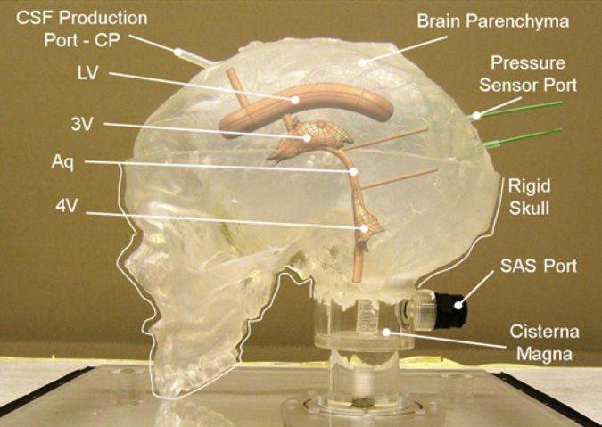
Supplementary Data

  • Article Type: Review Article
This is a required field
Please enter a valid email address
Approval was a Success
Invalid data
An Error Occurred
Approval was partially successful, following selected items could not be processed due to error