Full text loading...
Liquid cell transmission electron microscopy (TEM) has attracted significant interest in recent years. With nanofabricated liquid cells, it has been possible to image through liquids using TEM with subnanometer resolution, and many previously unseen materials dynamics have been revealed. Liquid cell TEM has been applied to many areas of research, ranging from chemistry to physics, materials science, and biology. So far, topics of study include nanoparticle growth and assembly, electrochemical deposition and lithiation for batteries, tracking and manipulation of nanoparticles, catalysis, and imaging of biological materials. In this article, we first review the development of liquid cell TEM and then highlight progress in various areas of research. In the study of nanoparticle growth, the electron beam can serve both as the illumination source for imaging and as the input energy for reactions. However, many other research topics require the control of electron beam effects to minimize electron beam damage. We discuss efforts to understand electron beam–liquid matter interactions. Finally, we provide a perspective on future challenges and opportunities in liquid cell TEM.
Article metrics loading...
Full text loading...
Literature Cited
Data & Media loading...
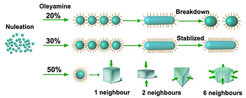
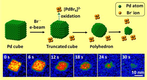
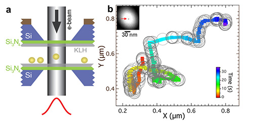
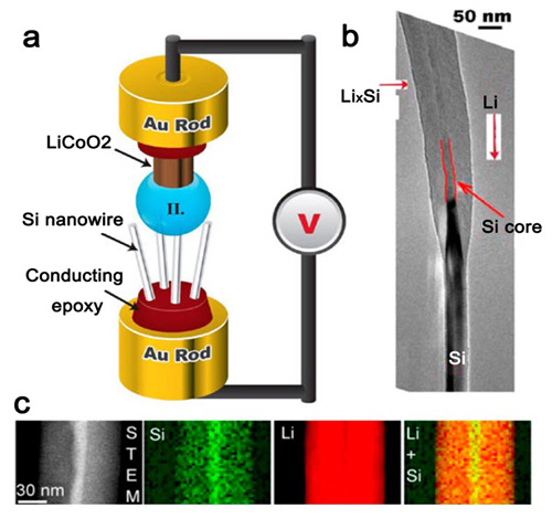
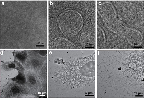
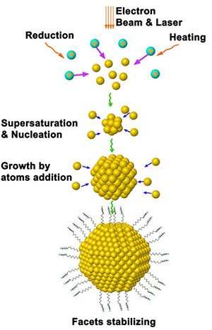
Download all Supplemental Figures as a single PDF, or see below: Supplemental Figure 1. A summary of oleylamine surfactant effects on platinum-iron nanoparticle shape evolution during growth. Figure reproduced with permission from Liao H-G, Zheng H. 2013. Liquid cell transmission electron microscopy study of platinum iron nanocrystal growth and shape evolution. J. Am. Chem. Soc. 135:5038—43. Supplemental Figure 2. In situ observation of platinum cube oxidative etching. Figure reproduced with permission from Jiang Y, Zhu G, Lin F, Zhang H, Jin C, et al. 2014. In situ study of oxidative etching of palladium nanocrystals by liquid cell electron microscopy. Nano Lett. 14:3761—65. Supplemental Figure 3. Manipulation and imaging of a gold nanoparticle movements in a liquid cell using an electron beam. (a) Schematic illustration of an experimental setup in which an electron beam passes through the silicon nitride window and traps gold nanoparticles inside the beam. (b) Trajectories of the electron beam movement and the global movement of the gold nanoparticle. Figure reproduced with permission from Zheng H, Mirsaidov UM, Wang L-W, Matsudaira P. 2012. Electron beam manipulation of nanoparticles. Nano Lett. 12:5644—48 and Zheng H. 2013. Using molecular tweezers to move and image nanoparticles. Nanoscale 5:4070—78. Supplemental Figure 4. (a) Schematic illustration of an open cell setup using an ionic liquid. (b) A silicon nanowire after lithiation. (c) Scanning transmission electron microscopy (STEM) image and electron energy loss spectroscopy map of the element distribution in the silicon nanowire after lithiation. Figure reproduced with permission from Gu M, Parent LR, Mehdi BL, Unocic RR, McDowell MT, et al. 2013. Demonstration of an electrochemical liquid cell for operando transmission electron microscopy observation of the lithiationdelithiation behavior of Si nanowire battery anodes. Nano Lett. 13:6106—12. Supplemental Figure 5. Imaging of wet biological samples by graphene liquid cell. (a) Low-resolution transmission electron microscopy (TEM) image of H3N2 viruses in a graphene liquid cell. (b) Circular virus showing a higher contrast for the outer membrane, which presumably indicates the presence of hemagglutinin and neuraminidase proteins. (c) Nonspherical viruses. (d—f) TEM images of MDCK cells with different magnifications. Figure reproduced with permission from Park J, Park H, Ercius P, Pegoraro AF, Xu C, et al. 2015. Direct observation of wet biological samples by graphene liquid cell transmission electron microscopy. Nano Lett. 15:4737—44. Supplemental Figure 6. The nucleation and growth of nanoparticles under an electron beam.